Image of a Capsule for Bacteria Clip Art of a Hot Springs
4 Bacteria: Prison cell Walls
Information technology is important to note that not all bacteria accept a jail cell wall. Having said that though, it is also important to notation that most bacteria (about 90%) accept a cell wall and they typically have one of two types: a gram positive cell wall or a gram negative prison cell wall.
The two dissimilar cell wall types tin be identified in the lab by a differential stain known every bit the Gram stain. Developed in 1884, information technology's been in use ever since. Originally, it was non known why the Gram stain allowed for such reliable separation of bacterial into two groups. Once the electron microscope was invented in the 1940s, it was plant that the staining difference correlated with differences in the cell walls. Here is a website that shows the actual steps of the Gram stain. After this stain technique is applied the gram positive bacteria will stain purple, while the gram negative leaner volition stain pink.

Overview of Bacterial Cell Walls
A cell wall, not merely of bacteria but for all organisms, is found outside of the cell membrane. It'southward an additional layer that typically provides some force that the cell membrane lacks, by having a semi-rigid structure.
Both gram positive and gram negative prison cell walls contain an ingredient known as peptidoglycan (also known equally murein). This particular substance hasn't been establish anywhere else on Earth, other than the cell walls of leaner. But both bacterial cell wall types contain additional ingredients as well, making the bacterial prison cell wall a circuitous construction overall, particularly when compared with the cell walls of eukaryotic microbes. The prison cell walls of eukaryotic microbes are typically composed of a single ingredient, like the cellulose plant in algal cell walls or the chitin in fungal cell walls.
The bacterial jail cell wall performs several functions likewise, in addition to providing overall strength to the jail cell. It also helps maintain the cell shape, which is of import for how the prison cell will grow, reproduce, obtain nutrients, and move. It protects the jail cell from osmotic lysis, as the cell moves from one surround to another or transports in nutrients from its surroundings. Since water can freely move beyond both the jail cell membrane and the cell wall, the cell is at gamble for an osmotic imbalance, which could put pressure on the relatively weak plasma membrane. Studies have actually shown that the internal pressure of a cell is similar to the force per unit area constitute inside a fully inflated car tire. That is a lot of pressure level for the plasma membrane to withstand! The jail cell wall can go on out certain molecules, such as toxins, particularly for gram negative bacteria. And lastly, the bacterial cell wall can contribute to the pathogenicity or disease –causing ability of the jail cell for sure bacterial pathogens.
Structure of Peptidoglycan
Let usa start with peptidoglycan, since information technology is an ingredient that both bacterial prison cell walls accept in common.
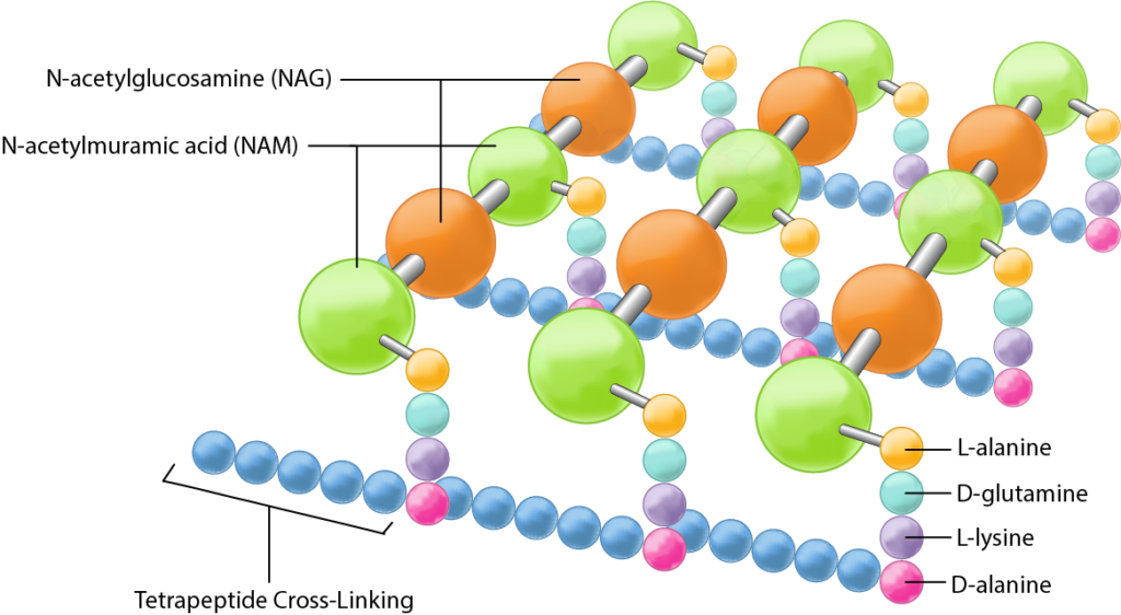
Peptidoglycan is a polysaccharide made of two glucose derivatives, N-acetylglucosamine (NAG) and Due north-acetylmuramic acid (NAM), alternated in long chains. The chains are cross-linked to one another by a tetrapeptide that extends off the NAM sugar unit of measurement, allowing a lattice-like structure to grade. The 4 amino acids that etch the tetrapeptide are: L-alanine, D-glutamine, L-lysine or meso-diaminopimelic acid (DPA), and D-alanine. Typically only the L-isomeric form of amino acids are utilized past cells only the use of the mirror image D-amino acids provides protection from proteases that might compromise the integrity of the cell wall by attacking the peptidoglycan. The tetrapeptides can exist directly cantankerous-linked to one some other, with the D-alanine on one tetrapeptide binding to the Fifty-lysine/ DPA on another tetrapeptide. In many gram positive bacteria there is a cross-bridge of five amino acids such as glycine (peptide interbridge) that serves to connect ane tetrapeptide to another. In either instance the cross-linking serves to increase the strength of the overall construction, with more strength derived from complete cross-linking, where every tetrapeptide is bound in some manner to a tetrapeptide on another NAG-NAM chain.
While much is still unknown about peptidoglycan, research in the past ten years suggests that peptidoglycan is synthesized as a cylinder with a coiled substructure, where each ringlet is cross-linked to the curlicue next to it, creating an even stronger structure overall.
Gram Positive Cell walls
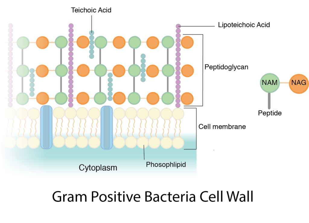
The cell walls of gram positive bacteria are composed predominantly of peptidoglycan. In fact, peptidoglycan tin can stand for up to 90% of the jail cell wall, with layer later layer forming effectually the cell membrane. The NAM tetrapeptides are typically cross-linked with a peptide interbridge and complete cross-linking is common. All of this combines together to create an incredibly potent jail cell wall.
The additional component in a gram positive cell wall is teichoic acrid, a glycopolymer, which is embedded within the peptidoglycan layers. Teichoic acrid is believed to play several important roles for the prison cell, such as generation of the net negative charge of the cell, which is essential for development of a proton motive forcefulness. Teichoic acid contributes to the overall rigidity of the cell wall, which is important for the maintenance of the cell shape, particularly in rod-shaped organisms. There is also evidence that teichoic acids participate in cell division, past interacting with the peptidoglycan biosynthesis machinery. Lastly, teichoic acids announced to play a part in resistance to adverse conditions such as high temperatures and high salt concentrations, as well as to β-lactam antibiotics. Teichoic acids can either exist covalently linked to peptidoglycan (wall teichoic acids or WTA) or connected to the jail cell membrane via a lipid anchor, in which instance it is referred to as lipoteichoic acid.
Since peptidoglycan is relatively porous, nigh substances tin pass through the gram positive cell wall with picayune difficulty. But some nutrients are too big, requiring the cell to rely on the use of exoenzymes. These extracellular enzymes are fabricated within the jail cell'south cytoplasm and and then secreted past the cell membrane, through the jail cell wall, where they function exterior of the jail cell to interruption down large macromolecules into smaller components.
Gram Negative Jail cell Walls
The jail cell walls of gram negative bacteria are more circuitous than that of gram positive leaner, with more ingredients overall. They do comprise peptidoglycan likewise, although only a couple of layers, representing 5-ten% of the total cell wall. What is most notable nigh the gram negative prison cell wall is the presence of a plasma membrane located exterior of the peptidoglycan layers, known every bit the outer membrane. This makes upwards the bulk of the gram negative prison cell wall. The outer membrane is composed of a lipid bilayer, very similar in composition to the prison cell membrane with polar heads, fat acid tails, and integral proteins. Information technology differs from the cell membrane by the presence of large molecules known every bit lipopolysaccharide (LPS), which are anchored into the outer membrane and project from the prison cell into the surround. LPS is made up of iii unlike components: 1) the O-antigen or O-polysaccharide, which represents the outermost function of the structure , two) the core polysaccharide, and 3) lipid A, which anchors the LPS into the outer membrane. LPS is known to serve many dissimilar functions for the prison cell, such as contributing to the internet negative charge for the cell, helping to stabilize the outer membrane, and providing protection from sure chemic substances by physically blocking access to other parts of the jail cell wall. In addition, LPS plays a part in the host response to pathogenic gram negative bacteria. The O-antigen triggers an allowed response in an infected host, causing the generation of antibodies specific to that office of LPS (think of E. coli O157). Lipid A acts equally a toxin, specifically an endotoxin, causing general symptoms of illness such every bit fever and diarrhea. A large amount of lipid A released into the bloodstream can trigger endotoxic daze, a body-wide inflammatory response which tin be life-threatening.
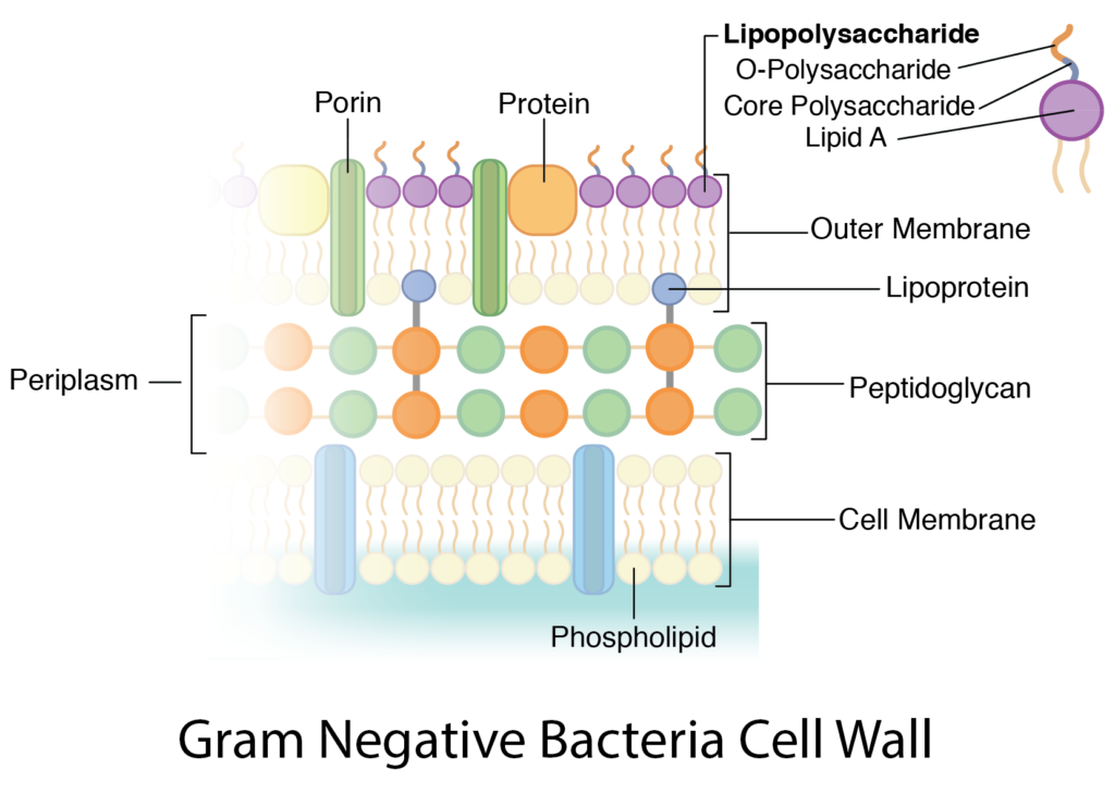
The outer membrane does present an obstacle for the cell. While there are certain molecules information technology would like to keep out, such every bit antibiotics and toxic chemicals, there are nutrients that information technology would like to let in and the boosted lipid bilayer presents a formidable barrier. Large molecules are broken downwardly by enzymes, in order to allow them to get past the LPS. Instead of exoenzymes (like the gram positive bacteria), the gram negative bacteria utilize periplasmic enzymes that are stored in the periplasm. Where is the periplasm, you lot inquire? It is the infinite located between the outer surface of the cell membrane and the inner surface of the outer membrane, and it contains the gram negative peptidoglycan. Once the periplasmic enzymes have broken nutrients down to smaller molecules that can go past the LPS, they still need to be transported across the outer membrane, specifically the lipid bilayer. Gram negative cells utilize porins, which are transmembrane proteins composed of a trimer of 3 subunits, which class a pore across the membrane. Some porins are non-specific and transport any molecule that fits, while some porins are specific and merely transport substances that they recognize by use of a binding site. Once across the outer membrane and in the periplasm, molecules work their fashion through the porous peptidoglycan layers before beingness transported by integral proteins beyond the cell membrane.
The peptidoglycan layers are linked to the outer membrane by the use of a lipoprotein known as Braun's lipoprotein (good ol' Dr. Braun). At 1 stop the lipoprotein is covalently bound to the peptidoglycan while the other end is embedded into the outer membrane via its polar head. This linkage between the two layers provides additional structural integrity and forcefulness.
Unusual and Wall-less Bacteria
Having emphasized the important of a cell wall and the ingredient peptidoglycan to both the gram positive and the gram negative bacteria, it does seem of import to point out a few exceptions likewise. Leaner belonging to the phylum Chlamydiae appear to lack peptidoglycan, although their cell walls have a gram negative structure in all other regards (i.e. outer membrane, LPS, porin, etc). It has been suggested that they might be using a protein layer that functions in much the same mode as peptidoglycan. This has an advantage to the cell in providing resistance to β-lactam antibiotics (such as penicillin), which attack peptidoglycan.
Bacteria belonging to the phylum Tenericutes lack a jail cell wall altogether, which makes them extremely susceptible to osmotic changes. They oft strengthen their jail cell membrane somewhat by the add-on of sterols, a substance usually associated with eukaryotic cell membranes. Many members of this phylum are pathogens, choosing to hide out within the protective environment of a host.
Key Words
prison cell wall, gram positive bacteria, gram negative bacteria, Gram stain, peptidoglycan, murein, osmotic lysis, N-acetylglucosamine (NAG), N-acetylmuramic acid (NAM), tetrapeptide, L-alanine, D-glutamine, 50-lysine, meso-diaminopimelic acrid (DPA), D-alanine, straight cross-link, peptide interbridge, complete cantankerous-linking, teichoic acrid, wall teichoic acid (WTA), lipoteichoic acid, exoenzymes, outer membrane, lipopolysaccharide (LPS), O-antigen or O-polysaccharide, core polysaccharide, lipid A, endotoxin, periplasmic enzymes, periplasm, porins, Braun's lipoprotein, Chlamydiae, Tenericutes, sterols.
Study Questions
- What are the basic characteristics and functions of the cell wall in Bacteria?
- What is the Gram stain and how does it relate to the different cell wall types of Bacteria?
- What is the bones unit structure of peptidoglycan? What components are present and how do they collaborate? Be able to diagram peptidoglycan and its' components.
- What is cross linking and why does this play such an important role in the cell wall? What different types of cross-linking are there?
- Why are D-amino acids unusual and how does having D-amino acids in the peptidoglycan keep this macromolecule stable?
- What are the differences between gram positive and negative organisms in terms of thickness of peptidoglycan, dissimilar constituents of PG and variations in cross linkage and strength, and other molecules associated with jail cell wall?
- What is teichoic acrid and what are its' proposed roles and functions? What are lipteichoic acids?
- What is the periplasm of gram negative bacteria? What purpose can it serve? What alternatives are available for cells?
- What is the general composition of the outer membrane of gram-negative microorganisms, its part and toxic backdrop? How is it linked to the jail cell? What is a porin and what are their functions?
- What grouping of bacteria lack peptidoglycan in their jail cell wall? What advantage does this confer?
- What group of bacteria unremarkably does non have cell walls and how do they maintain themselves?
Exploratory Questions (OPTIONAL)
- How does the mechanism of the Gram stain relate to specific components of the bacterial cell wall?
raiwalasputhessir.blogspot.com
Source: https://open.oregonstate.education/generalmicrobiology/chapter/bacteria-cell-walls/